Tick-borne Disease
Ticks can carry a number of organisms which can cause disease in dogs and people. Generally the organisms develop in the tick and are transmitted in the saliva as the tick feeds. There are six major groups of diseases we will briefly look at.
- Borreliosis (Lyme Disease)
The infectious agent is a spirochete (motile bacteria), Borrelia burgdorferi, which is carried by the deer tick, Ixodes spp. The white-footed mouse is the main reservoir for the spirochete and the host for the nymphal and larval forms of the tick. White-tailed deer are the definitive host with dogs and people being exposed to the adult ticks in deer- endemic areas. The lameness primarily presents due to joint pain and may be acute or chronic. It is often progressive. Signs of systemic disease include fever, loss of appetite, Lethargy and enlarged lymph nodes. The joints may be swollen. Radiographs show little evidence of degenerative joint disease. Blood work is often normal and Lyme blood titers are useful in unvaccinated dogs. Treatment consists of antibiotics and non-steroidal anti-inflammatory drugs for joint pain. A more unusual presentation seen primarily in the retriever breeds is a glomerulonephritis caused by the bacteria which can be fatal. - Ehrlichiosis
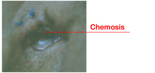
Ehrlichia spp. are rickettsial organisms which are intracellular parasites that replicate in host cells such as red blood cells, various types of white blood cells and platelets. Tick transmission by the brown dog tick, the lone star tick and the deer tick have been demonstrated. The parasitized cells are subject to early destruction in the spleen and liver as well as being disrupted by the life cycle of the parasite itself. Clinical signs are fever, lethargy, loss of appetite, weight loss, enlarged lymph nodes, and enlarged spleen. Often evidence of a bleeding disorder is seen with a bloody nose, bruising of the skin and small hemorrhages on the gums and other mucous membranes known as petechia. Inflammatory changes in the eyes can be seen as well. Arthritis in several joints, peripheral edema and muscle inflammation with pain are other recognized signs. Laboratory findings include low red cell counts, white blood cell counts, and low platelet counts depending on the species of Ehrlichia the dog is infected with. It is also common to see an increase in the serum proteins with protein loss in the urine. Abnormal blood clotting tests can be seen as well. The diagnosis is based on detecting the organisms within cells and serological testing for titers to the organism. Treatment usually consists of doxycyline as an antibiotic. If the disease is not detected early, recovery might take many months and the dog’s response may be poor or incomplete. - Rocky Mountain Spotted Fever
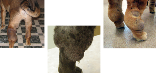
The cause of Rocky Mountain Spotted Fever is another rickettsial organism known as Rickettsia rickettsii and transmitted in the saliva of the American dog tick, the Lone Star tick, the Deer tick, the Rocky Mountain wood tick and the Pacific Coast tick. The disease name is a misnomer as the disease is found throughout the United States. Common signs include fever, lethargy, and loss of appetite. Skin signs are also common and include cutaneous edema, vesicles (blisters), bruising, and areas of skin sloughing. Other signs include joint pain and lameness, eye and nose discharge, coughing and breathing problems. Often there is anemia, low white blood cell counts and low platelet counts. Coagulation problems can develop. There can also be increases in liver tests and muscle enzyme levels. Serology is useful to diagnose the disease. Antibiotic treatment and supportive care are indicated. - Babesiosis
This disease is caused by a protozoan parasite (one celled organism) called Babesia gibsoni or Babesia canis. Transmission occurs via the brown dog tick and the organism invades the red blood cells resulting in severe anemia and systemic signs. Signs include hemolytic anemia caused by red blood cell destruction, fever, loss of appetite, weakness, and possible icterus (yellow color to the whites of the eyes and skin). It seems to be especially prevalent in Greyhounds. The major laboratory findings are the anemia or low red blood cell count, as well as low platelet count. The organism can sometimes be seen in the red blood cells under the microscope. Diagnosis is based on finding the organism and serological testing to detect antibody titers. Treatment involves specific drug therapy and supportive care. - Tick Paralysis
Ticks can inject a toxin when they feed. Most cases of tick paralysis involve the Rocky Mountain wood tick and the American dog tick. The toxin causes an ascending paralysis. Removal of the tick often reverses the signs. Death can result if the respiratory muscles are involved. - Bartonellosis
This is the group of organisms which can cause cat-scratch fever in man. The disease in dogs is transmitted by ticks. Bartonella are presumed intracellular bacteria which probably infects endothelial cells (cells that line blood vessels) and macrophages (cells which digest foreign materials). Signs may be inapparent or severe due to bacterial endocarditis and bacterial blood infection. Diagnosis is based on serology. Antibiotic treatment is not proven effective in clearing the infection. Not much is known about the disease in dogs at this time. A brief word needs to be mentioned regarding testing for these organisms. The use of blood serology detects the presence of antibodies to the disease-causing organism in the blood. Serology is circumstantial evidence in the courtroom of disease recognition. The presence of antibodies doesn’t really differentiate between exposure to the disease and actually having the disease. The presence of a positive serological test and clinical signs consistent with the disease is a strong case pointing to active infection. Ideally a blood test is done when the disease is first suspected and then a convalescent sample is taken 3-4 weeks after treatment. If the convalescent titer is much higher than the pretreatment titer it is felt the animal had an active infection and the body responded by increasing the antibody response. More recent advances in genetic testing have resulted in the PCR test or polymerase chain reaction test. In this test an actual DNA sequence of the disease-causing organism is identified which is conclusive proof that the organism is present and is causing disease. PCR testing can be done on a blood sample but can also be done on tissue harboring the organism.
We need to mention the importance of preventing tick exposure.
- Limit exposure – Ticks tend to favor tall grass, low brush areas and woods. Avoiding these types of terrain is best but keeping the dog on leash would suffice as well. If your property abuts areas of forest preserves, etc. fencing off the yard would be useful to limit exposure. Keeping high brush areas mowed or otherwise kept low is another approach. House infestations can result from bringing firewood into the house.
- Treat your yard – There are commercially available yard sprays containing permethrin which kill ticks on contact and degrade fairly quickly. Follow the manufacturer’s directions. Most are very safe once they dry. Local pesticide companies are another source of products and services.
- Tick control on pets – Topical products such as K9 Advantix™, Frontline TopSpot® and various flea and tick collars are effective to control ticks on animals. My favorite is K9 Advantix™ for Shar-Pei as it controls fleas, ticks and is a mosquito repellant which makes it useful in preventing West Nile disease. K9 Advantix™ CANNOT BE USED IN CATS. Also be careful to read the label carefully when purchasing insecticides from pet stores. Many products for dogs and cats look alike. I’ve seen several cases of dog products being used on cats resulting in toxicity.
- Vaccination – Dogs can be vaccinated to prevent Lyme’s disease. I currently only vaccinate those dogs at risk such as dogs with tick exposure and those that go on family vacations to Wisconsin and northern Illinois. Certainly field dogs, search & rescue and agility dogs may also fit in this category.
To remove a tick from your dog just grasp the tick with tweezers close to the skin and pull it out. It is a very rare occurrence to leave the head in. Disinfect the area with alcohol or antibacterial skin cleanser. There will be a noticeable reaction at the bite site for several weeks due to a reaction with the tick saliva and irritation. Also be careful not to get the blood or body fluids from the tick on your hands. That’s why it’s best to use tweezers or wear vinyl gloves when removing ticks. There is an infection potential if these fluids get into an open wound on your hand or finger. Ticks can be killed by placing them in alcohol. The diseases we talked about can be transmitted to people and cause similar diseases. Tick control in humans involves the use of insecticides, proper clothing and avoidance.
Due to the lack of specific symptoms with tick-borne diseases a high index of suspicion must be maintained. The turn around time on the serological tests is often 5-7 days or longer so I will often order those tests early in the workup for a sick dog. Antibiotic therapy is often started early as well because there is often a dramatic response to these drugs in dogs with tick- borne disease. Of course the best treatment is prevention.