Newsletter: Volume 7, Issue 3 December 2003
From Dr. Vidt…
I recently attended the AKC Canine Health Foundation Parent Club Health Conference held in St. Louis, MO in October. At the conference were representatives of various breed club health committees, members of the AKC Health Foundation and researchers, veterinarians and canine nutritionists involved in all aspects of canine health. Presentations covered canine cancer, canine nutrition, the Canine Genome Project, the use of breed health surveys, funding of canine health research, and the AKC Canine Health Information Center. It was a great time for me, as the Chairman of the Health Through Education Committee of the Chinese Shar-Pei Club, to meet with other breed health committee representatives, researchers and veterinarians.
Earlier this year at the Shar-Pei National Specialty held in Houston, TX in May, I was honored by the Chinese Shar-Pei Club of America with a Life-time Membership. It was a very unexpected and humbling gesture by the club and I will never forget their kindness.
Lastly, in this issue a new column by a Shar-Pei friend of mine begins. Sophie will have an advice column answering questions from other Shar-Pei and their owners. Should be interesting!
I hope everyone had a joyous Christmas season remembering the true meaning of Christmas and are looking forward to a Happy New Year spent with you Shar-Pei companions!
Dear Sophie:
I’m having an ongoing discussion with my owner about how often I need to take a bath. What are your thoughts. Kato
Dear Kato: I’m glad you asked that question. I usually take a bath when I get dirty or you can smell me walk into the room — that usually is every 4-6 weeks in the spring, summer and early fall. In the winter I don’t bathe as often. Any pet shampoo works well for general use. Different types of medicated shampoo may be prescribed by you veterinarian for different skin conditions and you veterinarian’s advice should be followed in using those. Bathing too often can result in dry skin problems or if you’re not dried thoroughly bacterial skin disease can result. Be sure you avoid getting water in your ears to avoid yeast ear infections. Keep soap out of your eyes also.
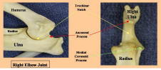
Elbow Dysplasia
The term “elbow dysplasia” encompasses a complex of orthopedic disorders involving the elbow joint of the dog. In order to differentiate among the different causes of “elbow dysplasia” a combination of diagnostic tools are used including physical examination, orthopedic examination, radiology, gait analysis, CT (computed tomography), MRI (magnetic resonance imaging), arthroscopy and exploratory joint surgery. Treatment options vary with the condition and will be discussed under the next section.
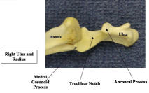
Components of elbow dysplasia:
1. Ununited anconeal process – The anconeal process is a small piece of bone making up the back part of the trochlear notch where the humerus articulates with the ulna. In the pup this process has a separate ossification center and usually fuses with the ulna by 4-6 months of age. If this process does not occur the fragment of bone will eventually result in secondary arthritic changes in the elbow joint. Clinical signs are usually seen in young dogs with intermittent or persistent foreleg lameness. When sitting or standing the dog tends to pull the elbow in and toe-out in the foot. Pain can often be elicited in the elbow on extreme flexion and/or extension of the joint. Treatment is the surgical removal of the fragment of stabilization of the fragment with a bone screw.
2. Ununited(Fragmented coronoid process – This condition is also known as fragmented medial coronoid process. Here the front part of the trochlear notch is involved – a small fragment of the medial coronoid process of the ulna is free in the joint and leads to development of secondary arthritis. Signs are similar to ununited anconeal process. Treatment is surgical removal of the fragment. This condition can be very difficult to diagnose with radiology and often the diagnosis has to be based on CT or MRI procedures.
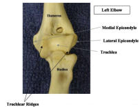
3. Osteochondritis dissecans of the medial or lateral humeral condyle – Osteochondritis dissecans (OCD) is caused by a defect in joint cartilage formation and maintenance that can manifest in several joint including the elbow. Typically a defect occurs in the joint cartilage leading to a flap of cartilage being raised or even breaking off into the joint space (a joint mouse). The clinical signs are the same as for the other elbow joint problems but the diagnosis is usually based on radiographs. Treatment is surgical management with removal of the cartilage flap or the joint fragment and debridement of the underlying bone to stimulate fibrous scarring of the defect.
4. Osteochondritis dissecans of the medial humeral trochlear ridge – The trochlea is the groove on the distal end of the humerus which articulates with the trochelar notch of the ulnar to form the hinge-like elbow joint.. On either side of the trochlea are the trochlear ridges. Cartilage defects can occur on the ridges leading to lameness. Surgery involves removal of the cartilage flap and debridement of the underlying bone.
5. Ununited medial epicondyle – This is an uncommon elbow condition characterized by detached calcified bodies just off the medial epicondyle of the humerus. They may or may not be associated with clinical signs such as lameness. Surgery may be needed in symptomatic cases.
The above conditions are typically seen in young, rapidly growing, large breed dogs indicating that rapid growth rate may be a predisposing factor. These conditions may also be related to trauma, a hereditary predisposition or nutritional factors.
Recommendations in Shar-Pei:
- Use of large breed puppy diets which are usually lower in protein and favor a slower growth rate. Avoid excessive weight in pups.
- Avoid trauma by supervising activity and exercise periods. Especially be careful with stairs, jumping up on or down from furniture, uncontrolled behavior, etc.
- Provide firm footing for puppies at all time.
Elbow incongruity – This refers to changes in the elbow joint configuration caused premature closure of ulnar and/or radial growth plates in pups. Growth plates, located at the ends of the long bones, allow for growth of the bone in length as puppies mature. Damage to these growth areas causes shortening of the limbs and places abnormal forces on the joint. This condition can occur in several locations in the front limb:
- Premature closure of the distal ulnar physis
- Premature closure of the proximal radial physis.
- Premature closure of the distal radial physis.Diagnosis is based on the combination of physical examination and radiographs. The orientation of the abnormal limb often indicates the region where the growth plate abnormality is located. Various surgical repairs are described but most involve an osteotomy (cutting the bone and removing a small section of it) of the bone in which the growth place has prematurely closed. This allows the normal bone to continue to grow in a normal manner. These problems are very often trauma related.
Elbow dysplasia appears to be increasing somewhat in frequency in Shar-Pei and quick diagnosis and early treatment can lessen the severity of arthritic changes in the elbow.
Click on picture to enlarge…